Diagnosing and Treating Keratoconus

A long-term study shows that about one in 2,000 individuals in the United States are diagnosed with keratoconus, an eye disorder marked by progressive thinning of the cornea. This condition causes the cornea to bulge in a cone shape and can cause problems such as blurry or distorted vision or sensitivity to light.
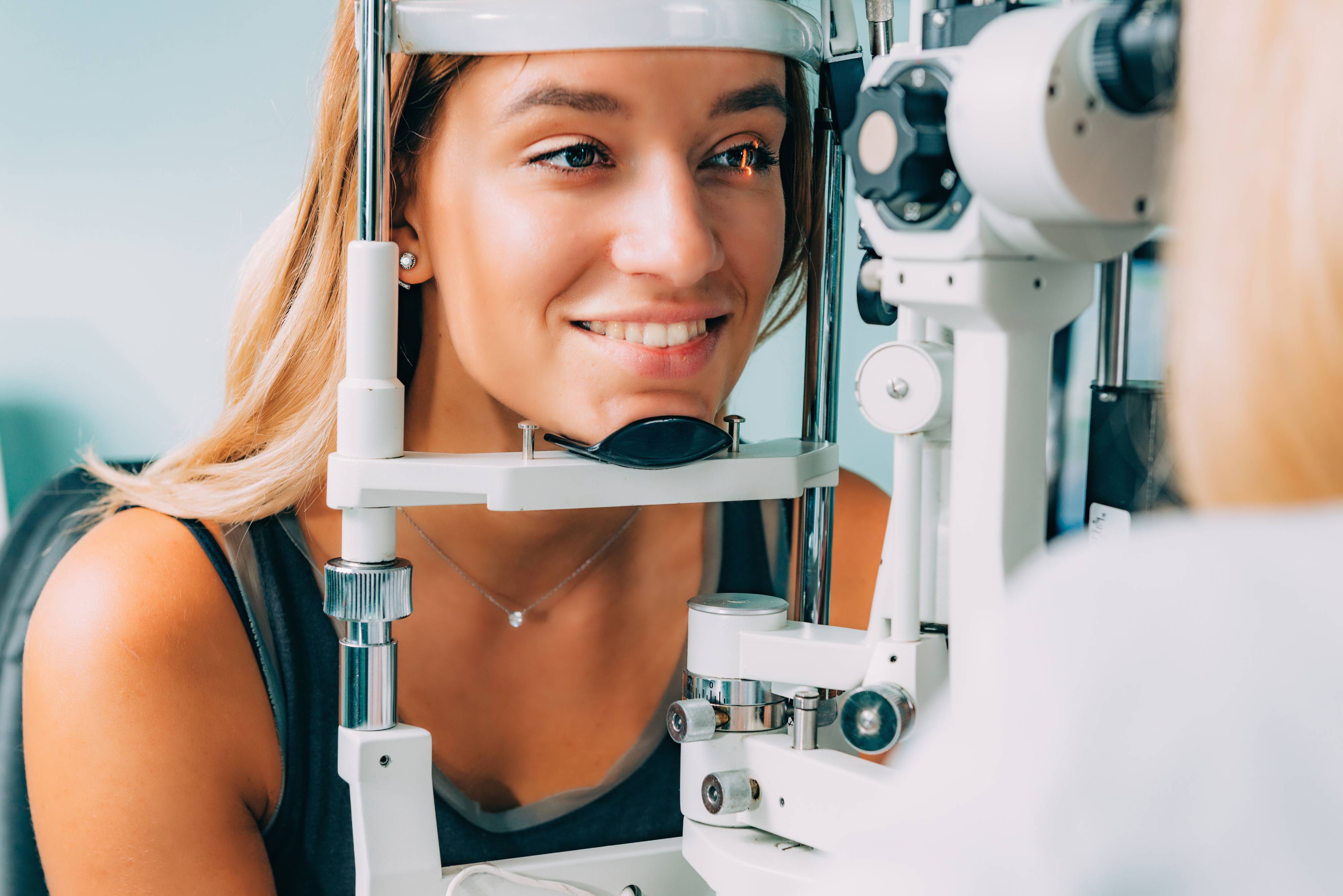
How Is It Diagnosed?
Corneal topography is the standard of care in detecting keratoconus. It is a simple, non-invasive process that lets your eye doctor examine the shape of your cornea. All you need to do when undergoing corneal topography is sit in front of a machine and look into a lens. A computer then records images of your cornea to create a detailed map that displays its shape and thickness. Corneal topography is the most accurate and one of the most common techniques in diagnosing keratoconus.
Keratometry is another method used to determine whether you have an eye disorder. It works by measuring the curvature and reflection of the anterior surface of your cornea. With this technique, your eye doctor will use a keratometer, a diagnostic instrument that shines a circle of light on your eye. This process allows your doctor to identify any irregularities in the shape of your cornea.
A more advanced form of topography, called tomographic corneal analysis, is now available. This process assesses both the cornea's front and back surfaces. This is crucial to early detection since the abnormalities found in keratoconus develop initially on the cornea's back surface. These abnormalities are often more severe than the irregularities on the front surface.
Your eye doctor may also use other diagnostic techniques to discover keratoconus progression at an early stage. These include aberrometry techniques and corneal optical coherence tomography, among others.
How Is It Treated?
Treatment for keratoconus generally depends on how severe your condition is and how fast the problem is progressing. Keratoconus often only has mild to moderate effects on your vision during its early stages. Your eye doctor will likely prescribe eyeglasses or soft contact lenses to correct your vision problems.
As your keratoconus advances, you will have to update your prescription time and again. If your disease progresses too much or too quickly, more specialized treatments may be necessary, such as:
Specialty Contact Lenses: If regular soft contacts don't work to correct your vision entirely, your eye doctor may recommend scleral lenses, hard contact lenses, piggyback lenses, or hybrid lenses.
Corneal Collagen Cross-Linking: This procedure can be completed in an invasive or non-invasive manner. It is designed to strengthen your corneal tissue and stop the bulging of your cornea as a result.
Corneal Inserts: Intacs® are small, transparent plastic devices that are surgically inserted into your cornea. These corneal inserts help flatten the curvature of your cornea to stabilize your vision.
Corneal Transplant: If your symptoms are severe enough, your eye doctor may suggest cornea transplant surgery. This treatment option is considered a last resort as it involves removing your cornea and replacing it with a donor cornea.
Early detection and intervention are crucial in managing keratoconus. Thanks to advances in technology, eye doctors can now diagnose and treat the condition more successfully. At iCare Vision Center, we can evaluate your symptoms and recommend the best treatment plan to restore your vision.
Learn more about diagnosing & treating keratoconus, contact iCare Vision Center in Seattle, WA at (206) 502-2800 to schedule your eye examination.


Contact Us
Clinic Hours
- Monday9 AM - 5 PM
- Tuesday9 AM - 5 PM
- Wednesday9 AM - 5 PM
- Thursday9 AM - 5 PM
- Friday9 AM - 5 PM
- SaturdayClosed
- SundayClosed
Closed for lunch from 1-2pm
Powered by: